SouthWest Breast Clinic
08 9711 1117

  • Home
  • Information for Patients
  • Information for GPs
  • Contact

The Triple Assessment

At the SouthWest Breast Clinic we know that every woman wants confidence in their results.  Whilst a single check can provide a good indication, the ‘triple test’ provides the highest level of confidence available and  is today recognised by medical practitioners around the world as the ‘gold standard’ diagnostic.  The triple assessment includes three diagnostic tests:

  • A review of your medical history and a clinical breast examination.
  • Medical Imaging – including a mammogram and/or ultrasound.
  • Pathology – on the cells collected from a fine needle aspiration (FNA) cytology and/or Core Biopsy.

 The sensitivity of the ‘triple test’ is greater than any of the individual components alone.

1. Clinical Assessment

Assessing your risk factors and examining your breasts

Assessing your risk factors and examining your breasts

Your breast physician will ask about your personal and family history of breast cancer, previous medical history and about any symptoms you may be experiencing.

They will then perform a clinical breast examination. This involves a thorough physical examination of the whole breast area, including both breasts, nipples, armpits and up to the collarbone.  

2. Radiology Assessment

Mammogram

Mammogram

We will then organise a mammogram for you at SKG Radiology, who are located directly across the road from the Breast Clinic. 

A screening mammogram is a low dose X-ray of your breasts, that can detect breast cancer at an early stage before it can be felt or noticed.  

Mammograms can often show a breast lump before it can be felt. They also can show tiny clusters of calcium called microcalcifications. Lumps or specks can be caused by cancer, fatty cells, or other conditions like cysts. 

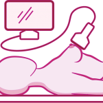
Ultrasound

Ultrasound

Depending on your personal circumstances (including your age and breast density) a breast ultrasound may be conducted (either before or after your mammogram).

Ultrasound uses sound waves to generate an image of the tissues inside the breast. A breast ultrasound can show all areas of the breast, including the area closest to the chest wall, which are hard to see with a mammogram.

This test is used to see whether a breast lump is filled with fluid (a cyst) or if it is a solid lump, which may require further testing.

3. Pathology Assessment

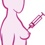
Tissue Biopsy

Tissue Biopsy

The final test we may request is a breast biopsy.  This test removes a small sample of tissue or sometimes fluid from the suspicious area. The removed cells are examined under a microscope and further tested to check for the presence of breast cancer. A biopsy is the only diagnostic procedure that can definitely determine if the suspicious area is cancerous.

The good news is that 80% of women who have a breast biopsy do not have breast cancer.

There are three types of biopsies:

Fine-needle aspiration

If the lump is easily accessible or we suspect that it may be a fluid-filled cystic lump, the doctor may choose to conduct a fine-needle aspiration (FNA). During this procedure, the lump should collapse once the fluid inside has been drawn and discarded. Sometimes, an ultrasound is used to help your doctor guide the needle to the exact site, whereby sound waves create a picture of the inside of the breast.

Core needle biopsy

This procedure involves the  removal of a small amount of suspicious tissue from the breast with a larger “core” (meaning “hollow”) needle. It is generally performed under local anesthesia . The radiologist will use specialized imaging equipment to guide the needle to the desired site where a tissue sample will be collected.

Symptoms of Breast Cancer

Related Posts

Warning Signs

Information for Patients

Symptoms of Breast Cancer

Breast Self Examination (2)

Information for Patients

How to do a breast self examination

Risk

Information for Patients

Calculating your risk of Breast Cancer

08 9711 1117

5 Ramsay St, Bunbury WA 6230
southwestbreastclinic.com.au